Friday, 31 July 2015

Pocket Friendly Cataract Surgery in Mumbai


More than 12000 cataract surgeries are done successfully has added charm to Krishna Eye Centre's achievements.
No Injection, No stitch, No patch Surgery.
Enjoy spectacle freedom with premium lenses.

cataract-services

When we look at any object, light rays from the object through the transparent cornea and the clear lens to strike the retina and form an image. The retina then relays the image to the brain through a set of wires called as Optic nerves.

What is Cataract?
For normal vision, it is imperative that the lens of the eye is clear and transparent, so that light rays can pass through it. When the lens becomes cloudy or opaque, light rays are not easily transmitted to the retina. This opaque lens is called as Cataract. Cataract usually develops gradually over a period and may affect both eyes in different degrees.

cataract

Ageing is the most common cause. As one ages, the protein in the lens break-down making it rigid and cloudy. Other than aging, many conditions can accelerate the break-down of the lens protein, like.
  • Diabetes
  • Eye Injury or prolonged exposure to radiation, sun or x-rays.
  • Smoking
  • Inflammation of the iris, uvea etc.
  • Disorders like hypothyroidism and atopic dermatitis.
  • Genetic diseases like Down's, Wilson's, Homocystinuria, Myotonic dystrophy etc.
  • Infections affecting the baby in the mother's womb like Herpes, Rubella, Syphilis, etc.
  • Medications like steroids and haloperidol.
How Does Cataract Affect Vision ?
Cataract develops very gradually and painlessly. It may affect one or both eyes.
  • Vision becomes Blurry as if looking through a frosted glass.
    blurred-vision 

  • Halos around object, especially bright lights e.g. Excess glare from oncoming headlights is noticed.
  • Loss of contrast sensitivity and dull appearance of colours.
  • Poor night vision.
  • Need for brighter light to carry out activities like reading.
Surgery alone is the treatment for Cataract. No medicine or diet can reverse Cataract formation. During the surgery the Cataractous lens is removed and is replaced by an artificial Intra Ocular Lens ( IOL ).
Phacoemulsification is the most advanced and accepted form of Cataract Surgery. During this surgery a very small opening of approximately 2-3mm is made in the eye and the cataract is removed by breaking it into small pieces, which are then gently sucked out with the same instrument. The incision taken is so small that it requires no stitches and heals rapidly. This makes it possible for the patient to resume normal activities early.

phacoemulsification

Once the Cataractous lens is removed, a new Intra Ocular Lens ( IOL ) is inserted in the same place.
There are a variety of IOLs which a patient can opt for. Typically there is a choice between foldable soft IOS and non-foldable hard IOS. Lenses could be either of PMMA, Silicon or Acrylic material. One can opt between Monofocal IOLs or Multifocal IOLs. The foldable IOLs are the most recommended. With monofocal IOL, the distance vision is clear without glasses but one has to wear reading glasses. With multifoal IOL, vision is clear both for distance and near and one is almost independent of glasses. 90-95% spectacle freedom is attained.

cataract-services

Toric IOLS are now available for patient with high cylindrical powers. Multifocal toric IOL can provide patients greatest level of spectacle freedom even if patients have a pre-operative corneal astigmatism.
Cataract surgery is done under topical anaesthesia. The new technique of eye drop anaesthesia (no injection) has distinct advantages over the conventional technique of giving injection anaesthesia.
In certain circumstances, the alternative technique of extra capsular cataract surgery with injection anaesthesia may need to be adapted as felt necessary for the best interest of the patient.
Cataract surgery has very predictable results with high success rate. The latest technique of NO INJECTION, NO STITCH and NO PATCH has made the surgery very easy for the patient; though it is more demanding on the surgical skills of your doctor. In some people natural capsule that support the IOL may became cloudy, several months after the surgery. If vision is affected, then laser can be used to open this capsule in a couple of minutes.

FAQs


When should surgery be done?
Cataract surgery is rarely an emergency surgery and should be considered when vision cannot be improved with new glasses or vision is no longer good enough for daily activities.

Which Lens Should on Select?
Each eye is unique. It is always good to customize the lenses best suited for the eye. Your Doctor will help you in selecting the lens.

Does The Artificial Lens Have An Expiry Date?
The artificial lens is designed to last for the rest of one's life. It needs to be replaced or adjusted only in cases of injury, severe reaction in the eye or dislocation which is very rare.

Do I need To Undergo Any Tests Before Surgery?
Yes. Routine Blood examinations are needed to rule out any infection in the body.
Well controlled Diabetes and Blood pressure are mandatory.

When Will I be Able to Resume My Activities After Surgery?
Most of the patients are back to their routine life in 2-3 days after surgery.


For online consultation with doctor & online appointments you can click on the following links : 

click
contactkrishnaeyecentre

Monday, 29 June 2015

Glaucoma Treatment in Mumbai at Affordable Cost

Glaucoma is a disease of the optic nerve. The optic nerve carries electrical signals from the eye to the brain and damage to it affects the vision. Glaucoma is usually a silent disease and also dangerous as the damage to the nerve is irreversible.

http://www.krishnaeyecentre.com/glaucoma.php

By the time a person realizes the symptoms, the disease is usually advanced. This makes early detection and treatment very important especially for older people.

http://www.krishnaeyecentre.com/glaucoma.php


Eye is like a ball. A clear liquid called as aqueous humor is constantly produced in very small quantities. An equal amount of this fluid flows out of the eye through microscopic drainage system. When this drainage system is blocked, excess fluid is retained, raising the pressure within the eyeball. The raised pressure pushes against the optic nerve and damages it. Sometimes there may be an acute attack due to sudden blockage causing a steep rise in the pressure with acute symptoms. This is an emergency.

People with glaucoma often do not have symptoms until vision loss occurs. When symptoms are present, they are :
  • Severe eye pain
  • Headache
  • Nausea / Vomiting
  • Rainbow Haloes Around Lights
  • Age above 40
  • Elevated eye pressure
  • Family history of glaucoma
  • High refractive errors
  • Past eye injuries
  • Thinner central cornea
  • Diabetes and migraine headache
  • Regular eye examination with routine measurement of intraocular pressure ( IOP ).
  • Optic nerve head evaluation.
  • Gonioscopy and ultrasound biomicroscopy ( UBM ) to check the drainage angles.
  •  Visual field analysis to check the function of the optic nerve.
  • Pachymetry for central corneal thickness
  • Newer imaging modalities like OCT, HRT and GDX may be required to document the anatomical damage to the optic nerve.
The treatment is aimed at arresting the progress of the disease. It is in the form of eye drops, laser and surgery.

http://www.krishnaeyecentre.com/glaucoma-services.phpThere are a variety of anti glaucoma medications available which have reduced the need for surgery. Some eye drops work to reduce the production of aqueous humor, whereas some work to open the drainage channels. Also combinations of medicines are available as eye drops for enhanced effect.

Laser Iridectomy ot Trabeculoplasty is helpful in certain cases to increase the drainage of fluid from the eye.

The surgical techniques of Trabeculectomy or modified Trabeculectomy with Mitomycin C are to be undertaken if medication is not able to arrest the progression of the disease.

New modality for control of glaucoma is the Express Shunt. Advanced Glaucoma might require the use of valve surgery to control the intra ocular pressure.

The purpose of treatment is to prevent further loss of vision. This is important because vision once lost due to glaucoma cannot be regained.

  • Always keep the medicine in stock before it gets over.
  • If more than one drug is used, wait for 10 minutes between dosages.
  • If one misses a dose, do not increase the number or amount of medication taken next time.
  • Do not stop taking medication while travelling.
  • Ask your doctor about how to use drops properly.
Don't Forget
  • Treatment for glaucoma requires a 'team' made up of both a compliant patient and doctor ( ophthalmologist ).
  • Doctor can prescribe treatment for glaucoma, but only patient can make sure to put eye drops regularly.
  • Do not change or stop taking medications without consulting the doctor ( ophthalmologist ).
  • Frequent eye examinations and test are important to monitor the eyes for any changes.

    Glaucoma generally has no signs or symptoms until serious loss of vision has occurred. The best way to protect the damage is early diagnosis, proper treatment and regular follow ups. Krishna Eye Centre provides glaucoma treatments at affordable cost.


    For more details contact us !
     Email : krishnaeyecenter@gmail.com 

    http://www.krishnaeyecentre.com/contact.php

Wednesday, 6 May 2015

PREMIUM LENSES USED IN CATARACT SURGERY

A cataract is clouding of the natural transparent lens of the eye which prevents the light rays passing through it thereby causing decrease in vision.


Why Surgery ?
Cataract can be treated only with surgery and no medicines can cure or prevent the cataract progression. Cataract Surgery is planned when one faces difficulties to carry out regular daily activities due to decreased vision which no longer improves with spectacles.
With the help of surgery the cloudy natural lens is removed and is replaced with a transparent artificial intra-ocular Lens (IOL) thereby improving vision.

What are the various Lens options ?
Choosing a correct IOL with regards to the design as well as the power forms the cornerstone of any Cataract surgery. To cope with the lifestyle changes, having good crisp vision has become a necessity. A correct premium IOL can provide one with independence from glasses. 

What are Monofocal IOLs ?
Traditional monofocal IOLs correct only the distant vision and for the near and intermediate activities patient needs to wear glasses.


What are Premium IOLs ?
The spectrum of Premium lenses includes Multifocals and Toric IOLs.
Multifocal IOLs
These IOLs consist of a pattern of concentric rings which helps focus the light falling onto the lens at different foci thus provide simultaneous near and far vision.
The different Multifocals available are
Bifocal IOLs :
These IOLs comprise of the concentric rings that focus the light passing through them at two points providing functional distance and near vision.


Imperfect intermediate distance vision clarity becomes a limitation with such lenses as intermediate distance vision is especially important during daily activities such as computer use, cooking and other such activities.

Trifocal IOLs : 
To overcome the limitation of the bifocal IOLs the newer concept of Trifocal IOLs was developed.
These lenses are designed in a way that they can provide good quality distance, intermediate and near vision simultaneously and one is almost independent of glasses. 90-95 % spectacle freedom is achieved.

What are Toric IOLs ?
Astigmatism is a condition where the cornea of one’s eye is more oval, like a rugby ball than the normal almost spherical shape. By the virtue of astigmatism one develops a cylindrical power of the eye.
This cylindrical power is not fully corrected with the traditional Monofocal or Multifocal IOLs and the patient requires spectacles for distance as well as near to overcome the blur caused by this kind of power even after cataract surgery in such cases.


A Toric IOL is premium IOL which can correct this cylindrical power as well and hence provide better vision quality when compared to the traditional non-toric options.

A Multifocal toric provides the greatest spectacle freedom

Monofocal Toric


Multifocal Toric
Benefits of Premium Lenses
  • Multifocal IOLs offer good vision not only at distance but also for near without the use of glasses or contact lenses as against the traditional monofocal lenses in which one requires to wear glasses for near activities.
  • Trifocal IOls provide a good quality vision simultaneously for near, intermediate and far distance giving patients freedom from eyeglasses.
  • Toric monofocal IOLs correct the cylindrical component of the eye power making the patients with astigmatism spectacle free for distant vision after cataract surgery.
  • Toric multifocal IOLs provide highest quality of spectacle freedom for patients with astigmatism.
For more details contact us..

Wednesday, 4 February 2015

Retinal Surgery in India

Retinal Detachment :

Retina is said to be a layer inside the eye which is light sensitive in nature. This is the layer which sends the visual messages to the brain through optic nerve. The retina is said to be detached when it is pulled or lifted from its normal position. Even vision loss can be caused due to retinal detachment if it is not treated properly.

Krishna Eye Center, Mumbai is one of the famous and well known hospital which provides best Retinal Surgery in India.

Types Of Retinal Detachment :

There are mainly 3 Types of retinal detachments. They are

  • Rhegmatogenous
  • Trectional
  • Exudative


Rhegmatogenous :

This is the most common type of retinal detachment. This is caused when a break or tear in the retina leads the fluid to get settled under the retina causing retinal detachment. Here retina get detached from its nourishing layer which contains a retinal pigment called as epithelium.


Tractional :

This is the less seen detachment. In this retinal detachment, the scar causes the retina's surface to contract, leading to detachment.

Exudative : 

This the the detachment which is caused due to retinal diseases including inflammatory disorders and trauma. This detachment is caused by the leaking of the fluid to the area under the retina. But there will not be any tears or breaks in the retina.

Risk Factors or Causes of Retinal Detachment :

There is no age for retinal detachment. It can be caused in any age. But there is more risk to happen in people who is aged more than 40.

The other causes includes,

  • People who are highly short sighted.
  • Having a family background of retinal detachment.
  • People who have undergone a cataract surgery in the past.
  • Having another eye diseases.
  • An early eye injury.


Symptoms of Retinal Detachment :

The main symptoms of retinal detachment includes

  • Appearance of floaters in the vision.
  • Light Flashes appearing in the vision.
  • Narrowing of vision.


Treatment :

  • At the early stages, small tears or holes can be treated by using lasik surgery and cryopexy.
  • Another option for retinal detachment is retinal surgery which requires the patient to stay in the hospital. 


For more information visit our site or contact us.
Website : www.krishnaeyecentre.com
Our mail id : krishnaeyecenter@gmail.com


Sunday, 21 December 2014

Effective Glaucoma Surgery in Mumbai

What is Glaucoma?

Glaucoma is a condition that causes harm to your eye's optic nerve and became dangerous when kept untreated. It is frequently connected with the development of  pressure inside the eye. Glaucoma has a tendency to be inherited and may not appear until the desired time.

The created pressure which is called as the intraocular pressure, can harm the optic nerve. This optic nerve is responsible for the transmission of images to the brain. If this damage to the optic nerve due to the pressure continues, the glaucoma can leads to the permanent vision loss of the patient. So without treatment glaucoma can lead to total blindness within a few years.

About 50% of the people with glaucoma doesn't show any early indications or pain due to this pressure. So every one should see an eye doctor regularly as possible since this can diagnose glaucoma at its early stage and thus can avoid the danger of losing the vision permanently.

If you are a diabetic patient or have an family history of glaucoma, then the chances for getting glaucoma is very high. So its better for those persons to undergo a comprehensive eye checkup.

Krishna Eye Center in Mumbai is one of the famous hospital providing high quality Glaucoma Surgery in Mumbai.


Causes of Glaucoma:


  • Glaucoma can be caused if you are aged over 40.
  • It can be caused as hereditary.
  • It can be caused due to diabetes.
  • It can also happen due to the use of steroids.
  • Due to any early damage to the eyes.


Symptoms of Glaucoma:


  • Visibility of halos while looking to lights.
  • Loss of vision.
  • Eyes colour changes to red.
  • Eyes became cloudy.
  • Vomiting tendency.
  • Eye pain.
  • Vision seems to be narrowed.


Diagnosis of Glaucoma:


  • Measuring the intra-ocular pressure by the method known as Applanation Tonometry.
  • Checking the corneal thickness by Pachymetry.
  • Optic disc is viewed by Ophthalmoscopy.
  • To evaluate the changes happened in the optic disc by using Fundus camera.
  • Mapping the functioning of optic nerve by the evaluation of the vision of each eye by using Visual Field Test.
  • To find out the early damages of the nerve fibers by using OCT.
  • Glaucoma type can be found out by using the Gonioscopy.


For more details about glaucoma CLICK HERE
Contact us by sending a mail or through our site..

Our mail id : krishnaeyecenter@gmail.com
Website : www.krishnaeyecentre.com


Saturday, 6 December 2014

High Quality Squint Treatment at Affordable cost in Mumbai

 What is Squint?

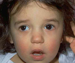
Squint is an eye disorder which results in the misalignment of eyes. Due to this problem both the eyes will not look like the same. Because of this there will occur other serious problems also. This may vary in accordance with the individuals. Now a days this disorder became common in children, but it is also seen in adults.


Krishna Eye Center, Mumbai which is having the best Hospital Infrastructure in Mumbai provides a high quality treatment for Squint at an affordable rate. This hospital is also famous for its well known Pediatric Ophthalmologist in India.

Squint in Children

Squint is now mostly seen in children


  • Squint in children is considerable only if the child is more than 2 months since the eyes of a newborn baby is rarely aligned during the first 2 months.
  • Squint damages the vision of the child.
  • Squint have to be treated at its early stage itself, otherwise it leads to losing of vision.
  • If the child grows with squint then regaining of lost vision become difficult.


What Leads to Squint?

The factors that may leads to Squint are


  • In some cases squint may occurs without any reasons.
  • Sometimes squint may occur hereditary.
  • Some infections in eyes may cause squint.
  • Other disorders like retinal diseases, tumors, glaucoma, cataract and optic nerve  diseases may sometimes leads to squint.
  • Due to weakening of eye muscles this misalignment takes place.
  • One of the major reason for squint is the abnormal impulses received by the eye muscles.
  • This may be also caused because of refractive errors.


Symptoms of Squint

The Symptoms shown by the effected persons are


  • Misalignment of the eyes.
  • Sometimes the effected ones will close the effected eye on looking to the sunlight.
  • In the case of children, they tilt and turn their heads for co-ordinating the vision through both eyes.
  • Double vision is another symptom shown by the effected persons in some cases.


Treatment for Squint

Squint can be treated according to the way of happening.


  • In some cases Squint will be due to refractive errors. In such case Non-surgical Treatment like use of spectacles is enough.
  • In other cases Surgical Treatment is suggested, which includes 3 steps : Vision Regaining, Alignment of Eyes, Binocular vision rectification.


For more information CLICK HERE
Contact us by sending a mail or through our site

Our mail id : krishnaeyecenter@gmail.com
Website : www.krishnaeyecentre.com


Saturday, 8 November 2014

Affordable Cost Eye Treaments for Children in Mumbai


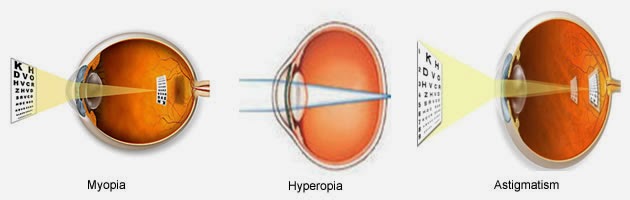
When light rays from a distant object do not come to a focus on the retina, the image of the object appears blurred and the condition is called Ametropis.

http://www.krishnaeyecentre.com/lasik.php

When the size of the eyeball is longer than normal, these light rays are focused in front of the retina. This is Myopia or short sightedness. This can be corrected by concave lenses as spectacles or contact lenses.
When the size of the eyeball is shorter than normal, these light rays are focused behind the retina. This is Hypermetropia or long sightedness. This can be corrected by convex lenses as spectacles or contact lenses.
A normal eye has spherical cornea in all its meridians. When the cornea is steeper of flatter in any of its axii it gives rise to Astigmatism. This is corrected by cylindrical lenses as spectacles or toric contact lenses.
Parents should encourage the sporting activities and provide a balanced diet rich in vegetables, milk products, salad, fruits and avoid junk food to maintain good eye health and overall health of their children.

http://www.krishnaeyecentre.com/paediatric-services.php
Amblyopia is commonly known as lazy eye. This should not be confused with squint. Amblyopia is poor vision in an eye that could not develop normal sight during early childhood.
At the age of eight years, the development of the part of the brain that processes vision is almost complete. If the brain has not received clear images from the weak eye, it starts neglecting this eye. The eye is then said to be amblyopic or lazy as the vision does not develop.

  • Refractive errors such as Short Sightedness, Long Sightedness or Astigmatism.
  • Large difference in spectacle powers between the two eyes.
  • Strabismus or squint.
  • Obstruction of vision by droopy eyelid, cataract or other diseases.
Management
Successful treatment depends on how severe the Amplyopia is and the age of the child when treatment begins. If the problem is detected early, successful treatment is possible and treatment time is shorter, if it is detected after the age of 8 years the success rate will be low.
..Spectacles
The child should be encouraged to wear his or her spectacles as much as possible throughout his waking hours if he or she is being treating for Amblyopia.
..Patching
The treatment consists of patching the GOOD EYE, so that the affected eye is made to work and the child is encouraged to see with it. It requires a tremendous amount of co-operation and understanding by the parents.
..Pharmacological treatment
Sometimes we prescribe some medications to help the amblyopic eye see well.

Squint or crossed eye is a condition where both eyes do not move together. One eye deviates either inwards, outwards, upwards or downwards while the other eye remains straight. It may also be alternating between the two eyes. Refractive errors are the most common causes of the squint, the other being lazy eye, nerve or muscle weaknesses and injury.
Management
http://www.krishnaeyecentre.com/squint.phpTreatment of squint requires a combination of glasses, exercises and surgery. Surgery may be needed in one eye or both eyes depending upon the squint, essentially it is a combination of loosing or tightening of eye muscles.
It is important to correct squint as it can lead to deep Amblyopia and psychological problems in children, loss of binocular vision, and loss of depth perception. Many simple activities like threading a needle, ability to understand the depth of staircase or liquids in a glass and functioning on a microscope become difficult.

Normally the tears are formed by the tear glands in the eyes and are drained off through two holes in the corners of the eyelids called punctae into the nose through a duct. The entire drainage system is called as Nasolacrimal duct system. Sometimes there can be a block at any level, at the punctum or in the ducts which may fail to open after birth resulting in persistent watering or discharge from the eyes. If left unattended this can lead to infection and painful swelling near the bridge of the nose.
Management
..Massaging
http://www.krishnaeyecentre.com/
70 to 80% of times the duct opens on its own time. Massaging the tear ducts is important to open the ducts. Massaging is done with the thumb or the forefinger, the stroke is to be firm and in the direction from the eyes to the nose downwards and inwards.
..Probin
If massaging fails a wire probe might need to be passed through the duct forcing it to open. This is done under anesthesia or sedation in small children.
..Lacrimal Duct Surgery
If the above methods fail to open the blocked ducts, surgery is recommended in children above 1 year of age. The surgery is done under general anesthesia.

Introduction
Cataract can affect a child as young as a new born baby. Cataract in children cause severe damage to the development of vision and needs to be treated as an emergency.
There are various causes of cataract in children like hereditary, genetic, infectious, metabolic, eye injuries etc. It is not possible to ascertain the cause in every child.
http://www.krishnaeyecentre.com/cataract.php
However, sometimes investigations are needed to find the cause as they have a big impact on child's life span and future quality of life.
What is the treatment for Cataract?
  • The treatment of cataract in a child is usually by surgery at the earliest.
  • Cataract surgery in a child is totally different as compared to a cataract surgery in adults. There are many issues that are to be dealt with in a different manner in terms of examination as well as surgeries.

http://www.krishnaeyecentre.com/diabetic-eye-treatment.php
ROP is a disease affecting the retina of premature infants. Very often premature babies are kept in tents of hyperbaric oxygen until they become fit to breath normally. The incidence of ROP is on a rise, this is due to improved survival rates of very low birth weight and extremely premature infants. The hyperbaric oxygen instigates the growth of abnormal vessels from the retina. The key disease element in ROP is growth of abnormal blood vessels with increased risk of bleeding or developing retinal detachment.
  • Which babies are at risk? Who should be screened?
The criteria for screening babies are based on two critical factors – the birth weight and the gestational age. In general infants with birth weight less than 1700 grams and gestational age below 34 weeks need screening.
  • When should screening be done? How frequently should the child be examined?
As a rule of thumb 1st screening should be over by Day 30 of baby's life. Very small babies (Weight < 1200 grams or < 24 weeks age) need to be screened within 2 weeks. Depending on the stage of the disease immediate treatment or repeat exams may be required.
  • What is the treatment for ROP?
Treatment is in the form of Retinal Lasers and surgery if Retinal detachment develops.


Krishna Eye Centre provides best treatment for your child.....

For more details our hospital, you can visit our site : www.krishnaeyecentre.com

http://www.krishnaeyecentre.com/
For other information you can directly contact us!!!
Email : krishnaeyecenter@gmail.com